
The spike glycoprotein of SARS-CoV-2 contains a cleavage site for host cell proteases called furins. Deciphering the role of this cleavage site during infection is important for understanding the origin of the pandemic virus and its disease pattern in humans.
Back in February it was not known if the furin site in the SARS-CoV-2 was cleaved by cell proteases, and whether its presence is required for infectivity. Both questions have now been answered.
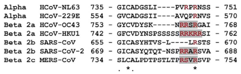
The figure shows amino acids at cleavage sites in the spike glycoproteins of various CoVs. A furin site is present in the spike glycoprotein of HCoV-OC43, HCoV-HKU1, MERS-CoV and SARS-CoV 2. It is called a multibasic site because it contains multiple basic (arginine) amino acids. The spike glycoproteins of HCoV-NL63, HCoV-229E, and SARS-CoV do not contain this multibasic cleavage site. Neither do SARS-related CoVs found in bats, including RaTG13, the virus with the closest overall genome sequence identity with SARS-CoV-2.
To study cleavage and function of the furin site, various CoV spike glycoproteins were engineered into vesicular stomatitis virus particles. This manipulation allowed the study of infected cells without the need for a BSL3 facility. The spike glycoprotein of SARS-CoV-2 was efficiently cleaved, while that of SARS-CoV or RaTG13 was not. Furthermore, when the SARS-CoV-2 furin site was exchanged with the corresponding sequence from SARS-CoV or RaTG13, no cleavage was observed. That cleavage was mediated by furins was verified by using specific protease inhibitors.
Cleavage of CoV spike glycoproteins is required for fusion of the viral and cell membranes upon entry. VSV harboring the spike of SARS-CoV-2 caused fusion of a human lung cell line; substitution of the furin cleavage site with the corresponding sequence from SARS-CoV or RaTG13 prevented cell fusion. However, VSV harboring the spike of SARS-CoV did cause fusion of these lung cells, due to cleavage by a different protease. These observations demonstrate that the furin cleavage site in the spike glycoprotein is essential for entry of SARS-CoV-2 into lung cells. In contrast, a monobasic cleavage site is sufficient for entry of SARS-CoV.
The activation of the spike glycoproteins of SARS-CoV-2 and MERS-CoV are therefore similar. They both must first be cleaved by furins followed by cleavage by a different cell protease, TMPRSS2.
An interesting question is the origin of the furin cleavage site it SARS-CoV-2. Its closest relative, the bat isolate RaTG13, does not have this site. Nor do any of the other bat SARS-like CoVs or the pangolin CoVs that have been isolated. However recently a newly isolated bat SARS-like CoV, RmYN02, was shown to contain a poly basic amino acid insertion in the spike glycoprotein. This observation supports the hypothesis that the furin cleavage site in SARS-CoV-2 arose by recombination among bat viruses in nature.
Pingback: SARS-CoV-2 furin cleavage site revisited – Virology Hub
Vincent Racaniello said:
> This observation supports the hypothesis that the furin cleavage site in SARS-CoV-2 arose by recombination among bat viruses in nature.
A recombination event is key to this hypothesis. In nature, it requires that a single animal was simultaneously infected by two viruses; a low probability but not implausible event. Recombination in the lab is also plausible but it requires Gain-of-Function type research on the RaTG13 virus. I have no idea how probable this lab scenario is but I’d prefer to see the lab hypothesis falsified rather than dismissed outright.
Bret Weinstein on his Darkhorse podcast thinks the lab hypothesis is much more likely but I think he is overly focused the respiratory infection of cave roosting bats rather than the gastrointestinal infection of bats that spread the disease to other animals through their guano outside of the cave environment.
Horseshoe bats practice “perch feeding” which means that nocturnal animals in the bat’s feeding ground are most likely to encounter infected guano; pangolins, civets, and racoon dogs fit the bill and are in high demand in the black market for exotic animals. The Mekong River basin is ground zero for this kind of doubly-infected recombination event.
Vincent – I don’t understand your logic here. You’re basically saying a virus that shares 61% similarity (RmYN02) is a smoking gun to a natural event simply because it also shares the insertion of multiple amino acids at the junction site of the S1 and S2? You realize this virus is something on the order of 11500 bp different from SARS-CoV-2…hardly the precursor virus.
Flyby News has posted a link to this page and to this presentation by Chris Martenson which supports the premise above. Martenson condenses his critique to a single question: “How did that polybasic furin cleavage site PRRA get into Covid 19?â€
Took a look at Flyby News. Appears to be a large scale compendium of conspiracy theories: JFK, 9-11, NASA, you name it.
E. Braun, D. Sauter. Furin-mediated protein processing in infectious diseases and cancer, August, 2019.
https://onlinelibrary.wiley.com/doi/pdf/10.1002/cti2.1073
Current review of (suddenly more important) therapeutic inhibition of furin.
The Göttingen paper confirms that without the furin clip, SARS-CoV-2 cannot infect human lung cells.
https://www.ncbi.nlm.nih.gov/pmc/articles/PMC5086990/
V. helpful PNAS paper from 2016 on the timing, sites and mechanisms of MERS infection of lung tissue. Note that for MERS, the furin protease not only enables lung cell infection — it puts it on a fast track. See Fig. 7, for example.
Hypoxia-enhanced Expression of the Proprotein Convertase Furin
Is Mediated by Hypoxia-inducible Factor-1
https://www.jbc.org/content/280/8/6561.full.pdf+html
There is a reluctance to use furin inhibitors as therapeutic agents because furin is a vital enzyme with hundreds of known substrates. However, when furin levels are much elevated, as they are in hypoxia, there might be some room to work.
This paper reports experiments in which the investigators determined that hypoxia, on average, tripled the expression of furin. Hypoxia-inducible factor-1 (HIF-1), a transcription complex, apparently produces the effect.
The advantage to SARS-CoV-2 seems clear. The furin clip is the rate-limiting step in the infection of human lung cells. As the infection progresses in the lungs, access to oxygen diminishes and furin becomes more and more abundantly available. A self-reinforcing spiral.
The authors also add some remarks about cytokines:
“…furin regulation by HIF-1 may be a more generalized phenomenon that could apply to other cellular contexts, including inflammatory condition, where furin activity was shown to be up-regulated (42).â€
Could a therapeutic target be HIF-1?
One of furin’s three promoters, P1, is known to be triggered by various cytokines, including interferon gamma, interleukins, and others. Net, there might be another self-reinforcing spiral involving cytokines.
Why hydroxyhloroquine didn’t work.
Chloroquine and hydroxychloroquine are thought to operate in the endolysosomal entry pathway into cells. Glance at the MERS infection entry pathways – early and late. The endosome pathway is a late entry pathway. SARS-CoV-2 probably doesn’t use it.
The furin clip enables the early action of a second protease, TMPRSS2, which in turn enables the virus to fuse directly with the target cell — and dump its RNA into the cytoplasm.
This is a shortcut past the late entry pathway, i.e., the endosome/lysosome pathway. Net, chloroquine and hydroxychloroquine work in a pathway that is simply bypassed — a direct consequence of the furin clip.
The TMPRSS2 inhibitor camostat is approved in Japan. Both camostat and the related agent nafamostat are in clinical trials as treatments for COVID-19 in the Netherlands and Germany. There are no approved furin inhibitors I am aware of, but one might ultimately think about a combination treatment including both furin and TMPRSS2 protease inhibitors.
Pingback: Computing Forever interview with Dolores Cahill contains numerous inaccuracies about COVID-19 and vaccines - Health Feedback
@John, potential explanations of the following observation?
Pseudovirus Entry of SARS-CoV-2 S-fur/mut (furin site removed) is 1.5-fold higher than SARS-CoV-2 S. See Figure 1A. ACE2 is a functional receptro for SARS-CoV-2 S protein. Entry of MLV pseudotyped with SARS-CoV-2 S, SARS-CoV S and SARS-CoV-2 S-fur/mut in VeroE6 cells. Walls et al., 2020, Cell 180, 281–292 Elsevier Inc. https://doi.org/10.1016/j.cell.2020.02.058.
The furin clip enables SARS-CoV-2 to infect human lung cells. It may infect other types of cells, possibly using other pathways and other clips at different rates.
The experiments described in Fig 1a of the Seattle paper targeted VeroE6 and BHK kidney cell lines from monkeys and hamsters.
The “… results suggest that S1/S2 cleavage during S biosynthesis was not necessary for S-mediated entry in the conditions of our experiments.â€
I don’t see a way to overlay these kidney cell studies on the processes of human lung cell infection by SARS-CoV-2, in which S1/S2 cleavage by furin is (we learned on April 28) essential.
The authors speculated that the value of the polybasic furin cleavage site in SARS-CoV-2 could be “to expand its tropism and/or enhance its transmissibility†over that of SARS. This turned out to be correct.
Can the furin cleavage insertion been accomplished using Crispr/Cas?
The following statement is false: However recently a newly isolated bat SARS-like CoV, RmYN02, was shown to contain a poly basic amino acid insertion in the spike glycoprotein. This observation supports the hypothesis that the furin cleavage site in SARS-CoV-2 arose by recombination among bat viruses in nature.
SARS-Cov-2 has a furin sequence P-R-R-A. The RmYN02 sequence has NO BASIN AMINO ACIDS!!!
The following statement is false: However recently a newly isolated bat SARS-like CoV, RmYN02, was shown to contain a poly basic amino acid insertion in the spike glycoprotein. This observation supports the hypothesis that the furin cleavage site in SARS-CoV-2 arose by recombination among bat viruses in nature.
SARS-Cov-2 has a furin sequence P-R-R-A. The RmYN02 sequence has NO BASIC AMINO ACIDS!!!
Vincent,
I am curious about the question from “Michael” on 23 May 2020 at 11:06 am:
“Can the furin cleavage insertion been accomplished using Crispr/Cas?”
Please see the following links? Perhaps they may help to shed light on an answer?
https://doi.org/10.1016/j.molcel.2019.09.013
https://zlab.bio/cas13
Pingback: The Evolution of Pandemics | FlybyNews
RmYN02 has not insertions at the S1/S2 junction. The alignment in the paper is wrong.