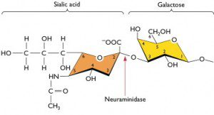

The cell receptor for influenza A virus strains is sialic acid. Human influenza A strains bind preferentially to sialic acids linked to galactose by an alpha(2,6) bond, while avian and equine strains prefer alpha(2,3) linked sialic acids (pictured). Alpha(2,6) linked sialic acids are dominant on epithelial cells in the human nasal mucosa, paranasal sinuses, pharynx, trachea, and bronchi. Alpha(2,3) linked sialic acids are found on nonciliated bronchiolar cells at the junction between the respiratory bronchiole and alveolus, and on type II cells lining the alveolar wall.
The 2009 swine-origin H1N1 influenza virus is known to bind both alpha(2,3) and alpha(2,6) linked sialic acids. This is consistent with the ability of the virus to cause lower respiratory tract disease. The D225G change might be expected to increase affinity for alpha(2,3) linked sialic acids. However, it is not known if increased binding affinity correlates with higher infectivity and pathogenicity. It’s equally likely that high affinity binding might restrict the movement of the virus in lung tissues by causing retention of the virus on nonsusceptible cells.
One view of the D225G mutation is that it is spreading globally and causing more severe disease. However there is no evidence in support of this hypothesis. According to WHO, viruses with the D225G change have been found in 20 countries since April 2009, but there has been no temporal or geographic clustering. As of January, the HA change has been identified in 52 sequences out of more than 2700. Furthermore, the authors of the Norwegian study write, “Our observations are consistent with an epidemiological pattern where the D225G substitution is absent or infrequent in circulating viruses, with the mutation arising sporadically in single cases where it may have contributed to severity of infection”.
One explanation for the sporadic emergence of influenza viruses with the D225G change is that they are selected for in the lower respiratory tract where alpha(2,3) sialic acids are more abundant than in the upper tract. Such selection might be facilitated in individuals with compromised lung function (e.g. asthmatics, smokers) or suboptimal immune responses, in whom the virus more readily reaches the lung. One way to address this hypothesis would be to compare the HA at amino acid 225 of viral isolates obtained early in infection, from the upper tract, with isolates obtained from the lower tract late in disease. However such paired isolates have not yet been obtained. But whether the presence of viruses with D225G increases viral virulence is unknown. Many H1N1 isolates from cases of fatal or severe disease do not contain this amino acid change.
There is an alternative explanation for the isolation of at least some influenza viruses with the D225G change: it is selected by propagation in embryonated chicken eggs. This selection occurs because cells of the allantoic cavity of chicken eggs have only alpha(2,3) linked sialic acids. A change in receptor specificity does not occur when viruses are propagated in MDCK (canine kidney) cells, which possess sialic acids with both alpha(2,3) and alpha(2,6) linkages. Consistent with this hypothesis, WHO reports (pdf) that the D225G substitution in 14 virus isolates occurred after growth in the laboratory.
Studies on the binding of influenza viruses to glycan arrays have shown that attachment is influenced not only by the linkage to the next sugar, but the type of sialic acid as well as the rest of the carbohydrate chain. The distribution of all the possible sialic acid containing sugars in the respiratory tract is unknown, as is the specific molecules that can support productive viral infection. The view that HA preferentially binds to either alpha(2,3) or alpha(2,6) linked sialic acids is likely to be overly simplistic: another casualty of reductionism.
Kilander A, Rykkvin R, Dudman SG, & Hungnes O (2010). Observed association between the HA1 mutation D222G in the 2009 pandemic influenza A(H1N1) virus and severe clinical outcome, Norway 2009-2010. Euro surveillance : bulletin europeen sur les maladies transmissibles = European communicable disease bulletin, 15 (9) PMID: 20214869
Takemae N, Ruttanapumma R, Parchariyanon S, Yoneyama S, Hayashi T, Hiramatsu H, Sriwilaijaroen N, Uchida Y, Kondo S, Yagi H, Kato K, Suzuki Y, & Saito T (2010). Alterations in receptor-binding properties of swine influenza viruses of the H1 subtype after isolation in embryonated chicken eggs. The Journal of general virology, 91 (Pt 4), 938-48 PMID: 20007353
Garcia-Sastre, A. (2010). Influenza Virus Receptor Specificity. Disease and Transmission American Journal Of Pathology DOI: 10.2353/ajpath.2010.100066

Pingback: uberVU - social comments
Pingback: The D225G change in 2009 H1N1 influenza virus | H1N1INFLUENZAVIRUS.US
Once again, your comments on D225G are quite misleading. The WHO data you are citing came out last year (in December – the WER you linked was just a repeat of the 2009 report) and was thoroughly refuted by sequences that were published in January (Russia and Ukraine in particular). The Ukraine sequences were by Mill Hill (deposited at GISAID) and demonstrated D225G, D225N, or both in 27/37 autopsy lung samples, clearly showing clustering in time and space (all samples were collected over a 2-3 week period in Ukraine) and all samples were phylogenetically similar. The Ukraine sequences were DIRECT and did NOT involve lab isolation. Moreover, like Norway, virtually all samples with D225G were from fatal or severe cases. The Norway study also noted that in four of their patients with D225G, samples from the upper and lower respiratory tract were available, and D225G was detected in both. The Norway sequences were from H1N1 isolated in MDCK (mammalian) cells and did NOT include egg isolates.
However, mixed infections with D225G, D225N, and wild type are common, so detection can be quite dependent on sample source, collection time, and lab propagation.
There are 5 HA sequences from autopsy lung from the 1918/1919 pandemic. Two have D225G, including the only sample from 1919.
In 1918/1919 tens of millions died and D225G clear transmitted.
Recent papers show the similarities between the receptor binding domain of the 1918 H1N1 virus and 2009 pandemic H1N1.
The importance of D225G has been OBVIOUS since the release of the initial sequences from Ukraine in November. Moreover, the frequent asscociation with wild type and/or D225N allows for transmission (as seen in two family members in Italy).
Human influenza virus prefers alpha(2,6), but chicken eggs have only alpha(2,3), then why human influenza virus can propagate in chicken eggs for vaccine production? Thank.
wo do have them 2,3 in the lower resp tract