
For my Ph.D. thesis project, I wanted to isolate reassortants of two influenza B virus strains, B/Lee and B/Maryland. The goal was to obtain viruses with a genome consisting of one RNA segment from one parent, and 7 RNA segments from the other parent. These viruses would then be used to identify the protein product of each viral RNA.
To isolate these reassortants, I co-infected cells in culture with both viruses, allowed them to replicate, and then harvested the newly synthesized viruses. I then did a plaque assay with the viruses produced by the co-infected cells, and isolated individual clones by plaque purification. I prepared virus stocks from each plaque-purified clone, and then asked if any of these viruses were reassortants.
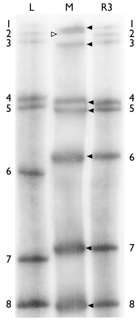
In 1979, we identified viral reassortants by a tedious method. Each virus to be examined was used to infect cultured cells in the presence of radioactive phosphate. The viruses were purified by centrifugation, and the viral RNA was extracted, concentrated, and fractionated by gel electrophoresis. The gel was dried and exposed to X-ray film. Because the viruses were propagated in the presence of radioactive phosphate, which was incorporated into the viral RNAs, it was possible to visualize each viral segment as a band on the film. The X-ray is shown above.
The RNAs of three different viruses are included: B/Lee on the left, B/Maryland in the middle, and a plaque-purified virus called R3. You can see that each lane contains 8 RNAs, as expected. It is also evident that the migration pattern of the RNAs is different for B/Lee and B/Maryland. This property allowed us to determine that the plaque purified virus R3 is a reassortant that inherits RNA 2 from B/Lee, and the remaining 7 RNAs from B/Maryland. Just what I wanted!
In fact, in just one infection, I isolated all the different reassortant viruses that I needed. I remember showing the gel to my thesis advisor: he told me I didn’t deserve such luck.
The whole procedure – from infecting cells in the presence of radioactive phosphate, to producing the X-ray, took about a week. And to get all the right viral reassortants required a great deal of luck.
Today, the process of identifying influenza viral reassortants is far simpler and faster. The process begins in a similar way – co-infect cells (or animals) with two different viruses. Once the infection is complete, a small sample of the cell culture medium is taken, heated to disrupt the virions, and the viral RNA is converted to DNA using reverse transcriptase. The DNA is amplified by PCR, in eight separate reactions, using primer pairs specific for the individuals segments. The products are then fractionated by gel electrophoresis, as shown here.
Total time to identify reassortants by PCR – less than a day. That’s progress.
In a few years, we’ll skip the gel electrophoresis and simply determine the sequence of the RNAs using a small, inexpensive machine that will be on most laboratory benches. And who knows what will be next? That’s one of the beauties of science: it is driven forward by technological innovation.
Racaniello VR, & Palese P (1979). Influenza B virus genome: assignment of viral polypeptides to RNA segments. Journal of virology, 29 (1), 361-73 PMID: 430594

I’m not entirely with you.
First, I only see 7 bands in the MD gel. And that 2 band in R3 looks a lot like L-3.
Not knowing anything about the virology and just looking at the gels, one might argue that R3-1 => L-2. R3-2 => L-3. And R3-3 => M-3, which would mean that R3 is lacking the 1 band and has 2 copies of the 3 band. Does the virology support this type of Ho? Do scrambled cultures drop segments out and/or double up on other segments?
Alternatively, if what we are seeing is M-1 and M-2 smeared, one could conclude the R3-1 and R3-2 are M-1 and M-2 properly separated (i.e. artifact). Is that the fast end of the gels at the top? Don’t you expect more wobble there?
As to bands 4/5, one interpretation is that R3-4 is common to all three gels and L-“4″ is really M-5, which in the Lee strain moves faster than the M-4 IOW, the 4/5 doublet could be flip-flopped in the Lee/Md strains. Do such doublets ever flip flop in these viral RNAs?
BTW, as these comments probably suggest, I am really jealous. At about the same time you were getting these beautiful results I was running gels (DNA) and getting spotted film that looked like shots from an X-ray telescope of galaxies in the deep universe! I knew I shoulda' gone into astronomy.
Some of the viral RNAs do migrate closely, and the photo doesn't help
by reducing the resolution. The first two RNAs of M (top of the gel =
slowest migrating, largest RNAs) run as a closely spaced doublet; as
we say 'it's evident on the original'. We did these gels multiple
times and in the end the segment assignments of the R3 virus were
convincing. They are run in 8M urea so everything is denatured and
running according to size.
The viruses do drop RNA segments and when they do, they are
noninfectious. If you have a high proportion of viruses lacking a
segment or two in any given viral stock, and then run a gel like this
one, you will see sub-molar ratios of specific RNA segments. Never
missing a segment – such a virus could not propagate on its own, e.g.
without a helper virus.
This gel system worked well, but it was finicky. Everything had to be
done just so, or else nothing. I had many smeared gels that never saw
the light of day.
Pingback: This Week In Virology « Influenza A (H1N1) Blog
Pingback: This Week In Virology – Esta semana en virologÃa « Gripe por A (H1N1) Blog