
In the summer of 1947 there were several small outbreaks of poliomyelitis in upstate New York. Gilbert Dalldorf, the director of the Wadsworth Laboratory in Albany, NY, and his associate Grace M. Sickles investigated this outbreak. In particular they sought polioviruses that could replicate in mice. This search was motivated by the fact that research on poliovirus required the use of monkeys which were extremely expensive. Dalldorf had attended the Fourth International Congress for Microbiology in 1947 where he heard that very young mice – suckling mice – could readily be infected with Theiler’s virus.
Dalldorf and Sickles made fecal suspensions from two children suspected of having poliomyelitis, and inoculated these into adult and suckling mice. Only the suckling mice (1 – 7 days old) developed paralysis; animals more than one week old were resistant to infection. The damage responsible for limb paralysis was widespread lesions in skeletal muscles, not in the central nervous system as occurs with poliovirus. Further study revealed that the viruses could be distinguished serologically from poliovirus.
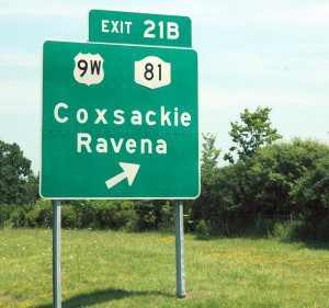
Not only had Dalldorf and Sickles identified the first members of a very large group of human viruses, but they also introduced and popularized a new and inexpensive animal into the virology laboratory – the suckling mouse. In 1949 Dalldorf suggested that the new viruses be called Coxsackie viruses, because the first recognized human cases were residents of that New York village. This unique name is of native North American origin.
Over ten years later the importance of this work was recognized by Dr. Max Finland of Boston City Hospital:
The isolation by Dalldorf and Sickles of viruses which produced paralysis with destructive lesions of muscle in sucking mice and hamsters, from the stools of two children with signs of paralytic poliomyelitis was an achievement that may rank in importance with Landsteiner and Popper’s production of human poliomyelitis in monkeys.
In subsequent years many different Coxsackieviruses were isolated that cause a variety of clinical syndromes. Today at least 30 serotypes of Coxsackieviruses are classified in the enterovirus genus of the Picornaviridae. The viruses are classified into groups A or B depending upon the pathological effect in suckling mice.
Not every locale is pleased to have a virus named after it. In May 1993, an outbreak of an unexplained pulmonary illness occurred in the southwestern United States, in an area shared by Arizona, New Mexico, Colorado and Utah called “The Four Corners.” Muerto Canyon was proposed as the name for the etiologic agent of the disease, because the virus was first isolated from a rodent near the canyon. However after residents objected, the name Sin nombre virus was given to the agent of hantavirus pulmonary syndrome.
Dalldorf G, & Sickles GM (1948). An Unidentified, Filtrable Agent Isolated From the Feces of Children With Paralysis. Science (New York, N.Y.), 108 (2794), 61-62 PMID: 17777513

Vincent, How did one specific virus – the typical childhood one that that gave my son blisters in his mouth and a high fever for three days – end up being known as Coxsackievirus, while the rest are not? T
The first Coxsackievirus was identified in 1947 in paralyzed children.
Subsequently about 29 more related viruses were identified that cause
a variety of clinical syndromes, including blisters and fever. Because
all 30 viruses were highly related, they were called Coxsackieviruses,
even though they were not isolated from that town.
Interesting, I didn’t realise the name was of native American origin. I became ill with ME/CFS after an outbreak of Coxsackie B4 in west of Scotland in 1983.
This one is very nice and excellent information and post of Exit.I like this Exit logo and information board design.I love this nice post.
exit signs
Better to post it here than on the XMRV posts…
From what I have read sofar, I think Enteroviruses are behind ME/CFS.First of all, there is the most recent work by Dr. John Chia. He finds VP1 enterovirus protein in about 80% of gut biopsies of people with ME/CFS he tested. Furthermore the viruses he find show about 80% homology to Coxsackie B.http://chronicfatigue.stanford.edu/infections/entero-experts.htmlThe work of John Chia is based on the earlier work of British scientists, mainly James Mowbray (if I’m not mistaken) who was the first to find VP1 in gut and muscle biopsies of ME/CFS patients.
But for me the key (and the best clinical description of the earlier outbreaks of ME/CFS I could find) is “The saga of Royal Free disease” by A. Melvin Ramsay: He describes how in villages in Icland in 1948 that suffered an ME/CFS outbreak were sparred from a following Polio outbreak that hit other villages. And he describes how an Polio outbreak in Adelaide ceased abruptly to be replaced by an ME/CFS outbreak.
Furthermore, if you haven’t read it, I can recommend the book Osler’s Web to get a feeling why ME/CFS is such a minefield – and a nightmare for those suffering from this disease.
GREW UP THERE THERE WAS AN OTHER OUTBREAK IN EARLY 50’S. 1953/54.
Pingback: Seven facts and a mystery about hand, foot and mouth disease | dreb
Pingback: PhD Seminar – Coxsackieviral Infection Susceptibility – HGSS McGill
Pingback: PhD Seminar – Susceptibility to Coxsackieviral Infection – HGSS McGill
Pingback: Day 362 (21q21.1-21q21.2): a Coxsackie virus interacting protein | Genome Year
Pingback: History of Coxsackievirus – Coxsackievirus