by Gertrud U. Rey
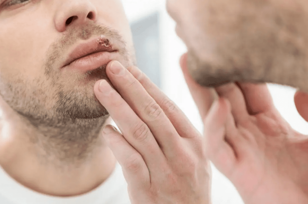
It is well known that stress and exposure to UV radiation can reactivate replication of latent herpes simplex virus type 1 and/or type 2 (HSV-1 and/or HSV-2), and the painful lesions associated with these infections. But why is that?


During a primary infection, HSV-1 and HSV-2 replicate within the epithelial cells of the skin and mucous membranes. Progeny viruses then travel to sensory neurons where they remain dormant as extrachromosomal “episomes” for the remainder of the host organism’s life. Occasional external stimuli can trigger their sporadic reactivation, potentially leading to renewed viral replication and symptomatic disease.
Many cellular functions are controlled by an assortment of externally triggered regulatory mechanisms that impact when and how certain genes are expressed, without changing the underlying DNA sequence. Such “epigenetic” regulations include processes like histone methylation and phosphorylation. Histones are proteins that function as spools around which DNA is wound into units called nucleosomes, which are then further coiled and condensed into a fibrous material called chromatin. Chromatinization protects DNA from damage, keeps it compacted, and prevents it from becoming tangled. However, this extensive coiling and folding also represses the expression of genes, because they cannot be accessed for transcription. The addition of methyl (CH3) groups to the histones can lead to further repression of gene transcription, depending on the location of the methyl groups. Further modification of histones by addition of phosphate groups (i.e., phosphorylation) can also prevent or induce the transcription of genes from that DNA, and hence, regulate the expression of these genes.
As it turns out, epigenetics plays a major role in the stress-induced reactivation of HSV. Latent HSV DNA is wrapped around histones that have three distinct methylation patterns, denoted as K9me2, K9me3, and K27me3. The methylated amino acid, lysine – labeled by its one letter abbreviation “K,” can be located at position 9 (i.e., K9) or 27 (i.e., K27) of the amino acid chain in the relevant histone. Additionally, K9 can be modified with either two or three methyl groups (represented as “me2” or “me3”). For example, the K9me3 modification has three methyl groups attached to the lysine at position 9 of the histone. Methylated lysines can bind additional proteins, which can further add to the repressive effects of methylation. In particular, K9me3 binds a transcriptional repressor known as heterochromatin protein 1, which blocks attachment of the factors needed to trigger a new round of replication and gene expression.
A pivotal study published in 2015 clarified the molecular mechanism by which stress induces reactivation of HSV-1 in mice. The authors showed that a kinase known as c-Jun N-terminal kinase (JNK) is essential for reactivating HSV-1. Kinases are enzymes that activate or inactivate functional molecules by phosphorylating them. The authors found that when they induced stress in mice with various drugs or stimuli, the amino acid serine (S10) located adjacent to K9me3 was phosphorylated, and this phosphorylation appeared to prevent binding of heterochromatin protein 1 to K9me3, thereby preventing its repressive effects. Phosphorylation of S10 was accompanied by reactivation of HSV-1, as evidenced by viral replication. The authors also found that when they induced stress while inhibiting JNK activity, S10 was not phosphorylated, and reactivation was blocked. These results confirmed that JNK-induced phosphorylation of S10 on chromatinized HSV-1 DNA is a crucial process in the reactivation of HSV-1. It is likely that JNK also phosphorylates the serine adjacent to K27 (i.e., S28), however, further studies are needed to confirm this hypothesis, and its relevance in reactivation of HSV-1.
It is noteworthy to mention that this mechanism was resolved in a mouse model, and it may not fully parallel what happens in humans. Nevertheless, these results provide valuable insights into the signaling pathways that appear to be key in HSV-1 reactivation, and thus warrant further exploration in the context of human infection. If JNK does reactivate HSV-1 in humans, a strategy to potentially prevent HSV-1/HSV-2 outbreaks in infected people could be to use drugs that block neuron-specific components of the JNK signaling pathway.
[The material in this blog post is also covered in Catch This Episode 60.]

All thanks belong to Dr Osato, the great herbal man that cured me from HERPES-1&2. I got diagnosed 7 months ago and I was desperately looking for a possible way to get this virus out of my body because I believe there is a cure somewhere. I keep searching until i saw people’s testimony about Dr Osato natural herbs on how he has being curing HERPES-1&2 with his natural herbs and i emailed him and tell him my problem and he prepare my cure and send it to me through UPS and gave me instructions on how to use the herbal medicine and behold after usage i went to the hospital for a checkup and the result was Negative and the symptoms of herpes was completely gone from my body which my doctor confirm it. You can contact Dr Osato on his website is https://osatoherbalcure.wordpress. com. Dr Osato cure so many different types of diseases/viruses with his natural herbs such as HERPES, HIV/AIDS, CANCER of all kinds, HSV 1&2, DIABETES, HPV and so many more. I want to thank God for using Dr Osato to cure me from genital herpes-1&2.
Any thoughts on how a virus parasite or microbial parasite causes brood parasitism (https://blogdredd.blogspot.com/2024/08/the-cuckoos-egg-hatched-again-4.html)?
And is any such suspect as adept as the HERPES perpetrators?
cf. https://blogdredd.blogspot.com/2024/08/the-cuckoos-egg-hatched-again-5.html
I was healed from HERPES through the help of Dr Grant. I saw a blog on how he cured people with his herbal medicine, I did not believe but i just decided to give him a try, I contacted him through his email and he prepared the herbal medicine for me which i took. after taking it 14 days, he told me to go for check up. could you believe that i was confirm HERPES negative after the test, and i went to a different hospital and it was also negative, I am so happy. If you are also infected with any disease like (1) HERPES,(2) DIABETES,(3) HIV&AIDS,(4) URINARY TRACT INFECTION,(5) HEPATITIS B,(6) IMPOTENCE,(7) BARENESS/INFERTILITY(8) DIARRHEA(9) ASTHMA… ETC kindly contact him now at grantingheartdesiresspell@ gmail. com and you can also call/text him on +2348115892498 or add Dr Grant on WhatsApp +2348115892498 and you will have a testimony… Good luck
All the symptoms of my genital herpes virus disappears totally after I finished drinking a herbal medicine from a herbalist so I went to several lab for test and they all came out Negative. Order and get Your herbal cure for herpes virus today from dr excel by visiting his website: https://excelherbalcure.com
I can proudly say now that I’m completely and permanently free from HSV (Herpes Simplex virus) I recently got in contact with a herbalist who prepared and sent me his herbal meds to drink which works magic on me. I went back for my tests at the lab and the doctor said I’m now negative after having the virus for years and all the obvious symptoms disappear immediately I started drinking the meds. May God continue to bless you Doctor Excel you are indeed a great herbal doctor. Contact Doctor Excel via his website: Excelherbalcure.com
I found out that herbal medicine is the best to get rid of herpes because I have just been cured from the virus, I took the healing process by contacting dr excel for natural treatment which really works wonders by neutralizing the virus, I have not feel the horrible symptoms and outbreaks anymore and my medical doctor told me the virus is gone after several test, I’m glad I finally got cured from this nasty disease. anyone with herpes should get in touch with this herbal man to get cured from that virus. his website: https://excelherbalcure.com