Although cancer therapies have improved dramatically in recent years, the main options for treating cancer still consist of surgery, chemotherapy, and radiation therapy. This limitation is a problem for aggressive cancers like glioblastoma multiforme (GBM), brain cancers which are typically resistant to traditional therapies.
GBM is a high grade glioma, an aggressive cancer of the central nervous system that is poorly immunogenic, meaning that it does not elicit a good host anti-tumor response. Because GBM tumor cells can migrate and invade normal brain tissue, the rate of relapse after treatment is high, and the average survival time after diagnosis is 12-16 months.
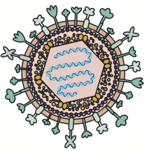
Oncolytic virotherapy is a therapeutic strategy that exploits the properties of human replication-competent viruses to infect and destroy cancer cells. Herpes simplex virus type 1 (HSV, pictured) is an attractive candidate for oncolytic virotherapy for several reasons:
- it is a prevalent human pathogen that does not integrate into the host DNA;
- it has a very large genome with many dispensable genes that can be replaced with foreign sequences;
- it infects a broad range of cell types and is naturally cytolytic, i.e., able to kill the cells it infects;
- it has fusion glycoproteins that can be engineered to specifically target cancer cells; and
- it is neurotropic, making it particularly suitable for treating GBM.
A research group at the University of Genoa in Italy engineered a replication-competent oncolytic HSV named R-LM113 to target the human epidermal growth factor receptor 2 (hHER2). This receptor is specifically expressed by GBM tumor cells and practically absent in healthy brain cells. Accordingly, R-LM113 is only able to replicate in cells expressing hHER2, and it is unable to enter cells expressing the HSV natural receptors. In a recent report, the same investigators further modified R-LM113 to include the gene for murine IL-12. IL-12 is a cytokine that increases the effectiveness of oncolytic viruses by recruiting T lymphocytes to initiate a cytotoxic response. The investigators then tested the efficacy of this IL-12-expressing virus (named R-115) in a GBM mouse model that is resistant to most cancer treatments.
To establish the mouse model, the authors infected neural precursor cells from BALB/c mice with a retrovirus encoding PDGF-B, a growth factor whose overexpression in these cells is known to induce gliomas. The cells were then transplanted into the brains of adult mice. Within about 100 days, the transplanted cells produced gliomas expressing markers that are histologically similar to human high grade gliomas and similarly poorly immunogenic. The authors harvested the gliomas, and then micro-dissected and cultured them to produce cells that they designated “parental tumor cells.” A portion of these parental tumor cells was engineered to express hHER2, generating “hHER2 tumor cells,” which were then transplanted into the craniums of mice, where they consistently produced high grade gliomas.
Three weeks after implantation of hHER2 tumor cells, different groups of mice were injected with the HSV constructs R-LM113, R-115, or a negative control treatment consisting of gamma-irradiated R-LM113. Control treatment recipients only survived an average of 13 days after receiving the treatment. Interestingly, while the median survival was similar for mice treated with R-LM113 and R-115 (33 and 35 days, respectively), there was a drastic difference in long-term survival between these two groups. Mice treated with R-LM113 all died within 48 days of treatment, while 27% of mice treated with R-115 were still alive 100 days after treatment. Two of these “long-term survivors” were euthanized and found to be tumor-free.
To see if R-115 induces a cancer-specific immune memory, two other R-115-treated long-term survivors received a second implant of hHER2 tumor cells, and despite the fact that they were not treated any further, they were still alive 220 days later. The authors then implanted these mice for a third time, this time with parental tumor cells lacking the hHER2 receptor. Remarkably, the mice still did not develop gliomas, suggesting that the initial treatment with R-115 elicited strong immunogenicity leading to long-term resistance to the tumor cells, even if the tumor cells did not express the hHER2 receptor.
Because they observed an increase in the number of T lymphocytes in the blood of mice shortly after infection with either R-LM113 or R-115, the authors analyzed the brains of mice for the presence of CD4- and CD8-positive T cells. This analysis revealed that the T cells tended to accumulate at the edge of the tumors in mice treated with R-LM113. In contrast, the CD4- and CD8-positive T cells deeply permeated the tumors in mice treated with mIL-12-expressing R-115. This observation suggests that mIL-12 may make the tumor microenvironment more permissive to the infiltration of cytotoxic lymphocytes.
It should be noted that treatment was timed to occur shortly before development of disease symptoms, based on previously determined survival data. It would be interesting to see if the treatment is still effective if it is initiated once symptoms are in full swing. Along these lines, it is reasonable to assume that the heterogeneity observed in response to treatment is attributable to differences in tumor sizes at the time of treatment. The percentage of infected cells in bigger tumors may be smaller than that in smaller tumors, a factor which may also affect the intensity of the immune response.
Although these results are encouraging, one must keep in mind that the mouse model doesn’t capture all the complexities of human cancers. As stated by Richard Klausner, former director of the National Cancer Institute, “The history of cancer research has been a history of curing cancer in the mouse. We have cured mice of cancer for decades – and it simply didn’t work in humans.” Nevertheless, mouse studies have yielded a wealth of information about human molecular biology and still provide an indispensable platform for preclinical research.
[Watch the video ‘Cancer Killing Viruses‘ for general information on viral oncotherapy – VRR]


Pingback: Cancer-killing viruses – Virology Hub