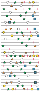

Single cells infected with vesicular stomatitis virus (VSV) were isolated using a glass microcapillary, and incubated overnight to allow completion of virus replication. Replication in a single cell imposes a genetic bottleneck, as few viral genomes are present. Virus-containing culture fluids were then subjected to plaque assay, during which 2 viral replication cycles took place. For each infected cell, 7-10 plaques were picked and used for massive parallel genome sequencing. A total of 881 plaques from 90 individual cells were analyzed in this way. Of the 532 single nucleotide differences identified, 36 were also present in the parental virus stock.
An interesting observation was that over half of the infected cells contained multiple parental variants. However, the multiplicity of infection (MOI) that was used should have only resulted in multiple infections in 15% of the cells. The results cannot be explained by RNA recombination as this process occurs at a very low rate in VSV-infected cells. The key is that MOI only describes the infectious virus particles that are delivered to cells. Because the particle-to-pfu ratio of VSV is high, it seems likely that many cells received both infectious and non-infectious particles. Furthermore, it is known that some RNA viruses may be transmitted to other cells in groups, either by aggregation of particles or within a membrane vesicle.
The conclusion from these results is very important: a single plaque-forming unit can contain multiple, genetically diverse particles. Plaque purification has been used for years in virology to produce clonal virus stocks, but at least for VSV, a plaque is not produced by a single viral genome.
The 496 single nucleotide changes that were not present in the parent virus arose after the bottleneck imposed by single cell replication. Between 0 and 17 changes were identified in the 7-10 plaques isolated from each cell. The single-cell bottleneck restricted the parental virus diversity to 36 nucleotide changes. In contrast, within 2 viral generations, the viral diversity was over ten times greater (496 changes). This observation illustrates the capacity of the RNA virus genome to restore diversity after a bottleneck.
The number of changes identified in the 7-10 plaques isolated from each cell, between 0 and 17, shows that some cells produce more diverse progeny than others. At least two sources of this variation were identified. The viral yield per cell varied greatly, from 0 to over 3000 PFU. Greater virus yields means more viral RNA replication, and more change for diversity. Indeed, greater virus yields per cell was associated with more mutations in the progeny.
Another explanation for the variation in single-cell diversity comes from analysis of cell #36. This infected cell produced viruses with 17 changes not found in the parental virus, more than any other cell. One of these changes lead to a single amino acid change in the viral RNA polymerase. This amino acid change appears to increase the mutation rate of the enzyme. Similar mutators – changes that increase the error frequency – have also been described in the poliovirus RNA polymerase.
RNA viruses must carry out error-prone replication to adapt to new environments. A consequence is that RNA virus populations exist close to an error threshold beyond which infectivity is lost. How the balance is maintained is not understood. The results of this study suggest that some infected cells may produce a highly diverse population, while in others a more conserved sequence is maintained. This distribution of diversity might permit the necessary evolvability without the lethality conferred by having too many mutations.
I would be very interested to know if the conclusions of this work would be changed by the ability to determine the sequences of all the viral genomes recovered from a single infected cell. The authors note that this is not technically possible, but surely will be in the future.

sounds very interesting – can you post a link to the paper?
The link to the paper is embedded in the third sentence of the first paragraph.
With respect to the last sentence: surely this IS possible these days? Even if done via cDNA PCR amplification?
Pingback: Viral variation in single cells | Cereal and gr...
The authors say it was technically not possible to sequence single cells. I’ve asked a few people who agree with this statement.