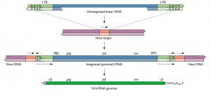

A search of the nucleotide sequence database with the previously identified XMRV integration site sequences revealed that 2 of the 14 sequences (from 2 patients) were identical to two XMRV integration sites in DU145 cells. This cell line was established in 1978 from the brain metastasis of a human prostate tumor. In early 2010 2007 DU145 cells were infected with XMRV, and sequences of two integration sites were determined (the database entries can be found here and here).
Identical retroviral integration sites have never been reported in independently infected cells. Furthermore, XMRV infection of DU145 cells was done in the same laboratory in which the XMRV integration sites were identified in prostate tumor DNA. The conclusion is that two of the 14 XMRV integration sites in prostate tumor DNA are likely to be the result of contamination. These prostate tumor DNA samples were probably contaminated with DNA from XMRV-infected DU145 cells.
These observations do not directly impugn the veracity of the other 12 XMRV integration sites identified in prostate tumor DNA. However, when DNA contamination occurs it is often ubiquitous. Hence the authors write:
Whilst it is conceivable that the other 12 integration sites apparently derived from prostatic tumor tissues are genuine patient-derived sequences, we suspect that some or all of them may also be the result of contamination with DNA from experimentally infected DU145 cells.
This possibility can and must be addressed experimentally.
Update: While writing this post I received an abstract from the 2011 Conference on Retroviruses and Other Opportunistic Infections (CROI) entitled “XMRV probably originated through recombination between two endogenous murine retroviruses during passage of a human prostate tumor in nude mice”. As usual I will await publication of this story in a peer-reviewed journal before discussing it further.
Garson JA, Kellam P, & Towers GJ (2011). Analysis of XMRV integration sites from human prostate cancer tissues suggests PCR contamination rather than genuine human infection. Retrovirology, 8 (1) PMID: 21352548
Stone, K., Mickey, D., Wunderli, H., Mickey, G., & Paulson, D. (1978). Isolation of a human prostate carcinoma cell line (DU 145) International Journal of Cancer, 21 (3), 274-281 DOI: 10.1002/ijc.2910210305
Dong B, Kim S, Hong S, Das Gupta J, Malathi K, Klein EA, Ganem D, Derisi JL, Chow SA, & Silverman RH (2007). An infectious retrovirus susceptible to an IFN antiviral pathway from human prostate tumors. Proceedings of the National Academy of Sciences of the United States of America, 104 (5), 1655-60 PMID: 17234809

In supplementary figure 3 of the Lombardi et al Science paper, the
authors show that a rat monoclonal antibody against SSFV env reacts,
in a western blot, with env of XMRV.
The monoclonal antibody to SSFV virus reacts with the SU protein of all MULVs, that was the point of using it. It demonstrated that the polytropic/xenotropic hybrid MLV class virus provisionally named as XMRV was a MLV virus.
That is not a cross reaction.
Lombardi et al consisted of four interelated but seperated experiments, and thus the results must be interpreted as a whole. When the serology experiments were carried out the virus had already been isolated and sequenced.
Thus when the combined results of the serology assays are looked at, then it is clear that the immune response was to a MLV class virus, which is what the serology assays were designed to show.
Fig. 4
Infectious XMRV and antibodies to XMRV in CFS patient plasma. (A) Plasma from CFS patients (lanes 1 to 6) were incubated with LNCaP cells and lysates were prepared after six passages. Viral protein expression was detected by Western blots with rat mAb to SFFV Env (top panel) or goat antiserum to MLV p30 Gag (bottom panel). Lane 7, uninfected LNCaP; lane 8, SFFV-infected HCD-57 cells. MW markers in kilodaltons are at left. (B) Cell-free transmission of XMRV to the SupT1 cell line was demonstrated using transwell coculture with patient PBMCs, followed by nested gag PCR. Lane 1, MW marker. Lane 2, SupT1 cocultured with Raji. Lanes 3 to 7, SupT1 cocultured with CFS patient PBMCs. Lane 8, no template control (NTC). (C) Normal T cells were exposed to cell-free supernatants obtained from T cells (lanes 1, 5, and 6) or B cells (lane 4) from CFS patients. Lanes 7 and 8 are secondary infections of normal activated T cells. Initially, uninfected primary T cells were exposed to supernatants from PBMCs of patients WPI-1220 (lane 7) and WPI-1221 (lane 8). Lanes 2 and 3, uninfected T cells; lane 9, SFFV-infected HCD-57 cells. Viral protein expression was detected by Western blot with a rat mAb to SFFV Env. MW markers in kilodaltons are at left. (D) Plasma samples from a CFS patient or from a healthy control as well as SFFV Env mAb or control were reacted with BaF3ER cells (top) or BaF3ER cells expressing recombinant SFFV Env (bottom) and analyzed by flow cytometry. IgG, immunoglobulin G.
This showed that the virus manufactured MLV class proteins.
“We next investigated whether XMRV stimulates an immune response in CFS patients. For this purpose, we developed a flow cytometry assay that allowed us to detect Abs to XMRV Env by exploiting its close homology to SFFV Env (16). Plasma from 9 out of 18 CFS patients infected with XMRV reacted with a mouse B cell line expressing recombinant SFFV Env (BaF3ER-SFFV-Env) but not to SFFV Env negative control cells (BaF3ER), analogous to the binding of the SFFV Env mAb to these cells (Fig. 4D and S6A). In contrast, plasma from seven healthy donors did not react (Fig. 4D and fig. S6A). Furthermore, all nine positive plasma samples from CFS patients but none of the plasma samples from healthy donors blocked the binding of the SFFV Env mAb to SFFV Env on the cell surface (fig. S6B). These results are consistent with the hypothesis that CFS patients mount a specific immune response to XMRV. ”
Well why was XMRV found and not found at various times in the Rhesus Monkey’s blood but always found in the tissues?
Brain Demyelination/lesions AIDS Yes ME/CFS Yes
Chronic sore throat Flu like illness AIDS yes ME/CFS yes
Swollen lymph nodes AIDS yes ME/CFS Yes
Cognitive problems AIDS yes ME/CFS yes
Skin Problems AIDS yes ME/CFS yes
IBS and other stomach problems AIDS yes ME/CFS Yes
Cancer/Leukimia/Lymphoma AIDS yes ME/CFS Yes
Weird tumor
spleen,liver,brain,testicular AIDS yes ME/CFS yes
Heart Failure AIDS yes ME/CFS yes
Many active viruses AIDS yes ME/CFS yes
White thrush AIDS yes ME/CFS yes
Swollen tongue with teethmarks around it AIDS yes ME/CFS yes
Fatigue AIDS yes ME/CFS yes
Seizures AIDS yes ME/CFS yes
How blind and gullible do you have to be, to not be able to see that ME/CFS is also caused by an HIV like virus? ME/CFS is caused by a retrovirus, we have known this since the early 80s when ME/CFS brain MRIs were taken and they looked exactly like AIDS patients, in the early 90s De Freitas found a retrovirus in the blood of ME/CFS patients and the CDC didn’t even bother paying attention to her, now the WPI has found the XMRV virus which is a retrovirus which was suspected all along and people still can’t admit that ME/CFS is caused by just that.,
How blind and dumb are you?
How can you tell me that is all in my head ?when two of my friends that have HIV have the very same health problems that I do but they do much much better then me because they have meds and I don’t.
They also don’t preferentially contaminate samples from patients with symptoms of illness…Â
But a scientist could preferentially contaminate patient samples (e.g. by not doing proper blinded studies). And I am using the term “scientist” very loosely here.
so we wait. Contaminants do not make antibodies.
And we will have to wait until Lipkin finds out if anybody can actually reliably distinguish patients from controls in a blinded fashion using said antibody tests.
As far as I know, Lombardi et al 2009 does not state that the tests were done blinded.